A week in which the radiologists made their case
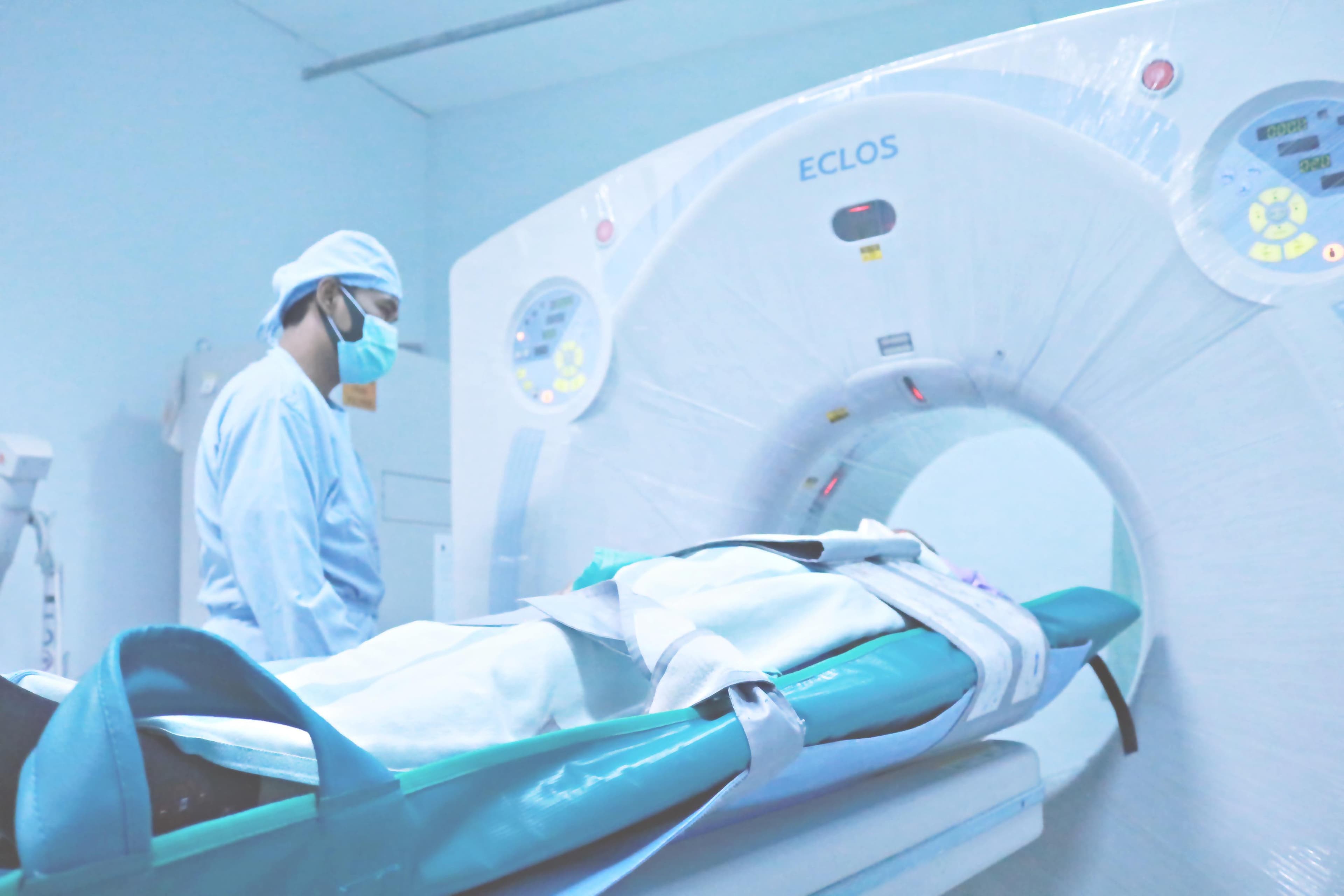
Every trauma trainee hears the line sooner or later, usually from someone senior and usually in the small hours: the CT scanner is the “doughnut of death”. Send a bleeding patient down the corridor into the gantry and you may get beautiful images of the injury that kills them while they are still inside it. The phrase survives because it encodes a real lesson. It also survives because nobody has quite agreed on when, or where, it stopped being true.
This week gave the argument fresh fuel. On 30 October the European Society of Emergency Radiology published new ESR Essentials practice recommendations on trauma whole-body CT (WBCT), led by radiologists at St Mary’s Hospital in London [1]. They describe WBCT as enabling rapid and comprehensive assessment of major trauma, and their first recommendation is that protocol selection should be tailored to haemodynamic status and mechanism of injury, not that some patients should never enter the scanner. The following day, Emergency Radiology published a review devoted to abdominal injuries missed on the initial trauma CT, with their pitfalls and mimics [2]. Put the two papers side by side and you have the modern paradox. The scanner now sits at the centre of the trauma pathway, yet it can still miss what matters.
ATLS candidates live inside that paradox. The course teaches that CT belongs to the haemodynamically stable patient, while the UK major trauma centre where you work may scan almost everyone within minutes of arrival. Which is right? The honest answer is “both, in different buildings”, and understanding why is worth more than either slogan.
What ATLS actually teaches
ATLS frames imaging in two tiers. Adjuncts to the primary survey (chest and pelvic radiographs, FAST or eFAST) are performed in the resuscitation bay without moving the patient. CT is an adjunct to the secondary survey, undertaken once resuscitation is established and the patient is physiologically ready to travel [3]. As a recent Swiss study summarised, ATLS recommends cross-sectional imaging with CT only in haemodynamically stable trauma patients; for those in shock, eFAST and conventional radiographs are the recommended imaging adjuncts [4]. The 11th edition’s xABCDE sequence makes the underlying logic even more explicit by putting control of exsanguinating haemorrhage first [5]. A patient who is bleeding to death needs the bleeding stopped, not a more elegant diagnosis.
That doctrine was written for every hospital in which ATLS is taught, including those with no scanner at all. It is a floor, not a ceiling. The question is what happens when the floor is raised.
How the pan-scan won the argument
The intellectual challenge came early and, fittingly, from London. In 2009 Otto Chan of the Royal London Hospital argued in the British Journal of Surgery that a primary CT survey should replace the primary clinical survey in major trauma, with the scanner sited so that resuscitation, surgery and interventional radiology could share one table [6]. The same year, a German registry study in The Lancet supplied the numbers. Of 4,621 blunt trauma patients, 32% had whole-body CT during resuscitation. Their standardised mortality ratio based on TRISS was 0.745, against 1.023 for those without: a 25% relative reduction in mortality (13% using the RISC score), with a number needed to scan of between 17 and 32 [7]. A 2014 meta-analysis of seven studies and 25,782 patients produced a pooled odds ratio for death of 0.75 in favour of WBCT [8].
England institutionalised the shift. NICE guideline NG39 recommends whole-body CT, from vertex to mid-thigh, for adults with blunt major trauma and suspected multiple injuries, and advises against using FAST or other imaging before immediate CT [9]. The Royal College of Radiologists’ 2024 adult trauma guidance goes further: contrast-enhanced whole-body CT is the default imaging procedure for severely injured patients, the scanner should be adjacent to or in the emergency room, and plain films of the abdomen, pelvis and spine are not required beforehand [10]. (We examined what this means for FAST in our earlier post on AI-assisted ultrasound. Here the question is the scanner itself.)
The trial that did not play along
Then came the randomised evidence, and it was awkward. REACT-2, a Dutch-led trial across five trauma centres published in The Lancet in 2016, randomised severely injured patients to immediate total-body CT or to conventional imaging followed by selective CT. In-hospital mortality was identical: 16% in each arm (86 of 541 versus 85 of 542), with no significant difference in the polytrauma or traumatic brain injury subgroups [11]. Immediate total-body CT did deliver answers faster, with a median time to diagnosis of 50 versus 58 minutes, but those eight minutes did not translate into lives saved [11, 12].
Supporters of WBCT have reasonable rejoinders. The trial took place in mature trauma centres where the conventional pathway was itself quick, so it compared two good systems rather than modern care with old-fashioned care. Independent appraisals also note that 46% of the control group ended up having every body region scanned sequentially anyway, which narrows the contrast considerably [12]. Even so, REACT-2 remains the best evidence we have: in a high-functioning system, scanning everyone immediately did not reduce deaths compared with scanning selectively. The 25% registry signal vanished under randomisation, a familiar story in trauma research, where the sickest patients are the least likely to reach the scanner in observational data.
From doughnut to hybrid room: the unstable patient
The more radical claim concerns patients in shock. ATLS keeps them out of the scanner; a growing body of observational work suggests that, in the right building, they may do better inside it.
A 2013 analysis of 16,719 blunt major trauma patients from the German TraumaRegister found WBCT independently associated with survival in both moderate shock (odds ratio 0.73) and severe shock (odds ratio 0.67), with a number needed to scan of 20 in severe shock [13]. The authors were careful: WBCT appeared safe and justified in unstable patients when performed quickly within a well-structured environment. A year later the same group showed that geography matters. Among 8,004 patients across 312 hospitals, a scanner inside the trauma room carried the lowest odds of death (odds ratio 0.68), while distances of more than 50 metres had a significant negative effect on outcome [14].
Japan took the logic to its conclusion with the hybrid emergency room, in which a sliding-gantry CT scanner, angiography and an operating table share a single space. At one Osaka centre, comparing 336 hybrid-room patients with 360 historical controls, median time to CT fell from 26 to 11 minutes, time to emergency procedure from 68 to 47 minutes, and the adjusted odds of 28-day death halved (odds ratio 0.50), with fewer deaths from exsanguination [15]. A 2024 Lausanne registry study of 161 patients in physiologically defined circulatory shock found 24-hour mortality of 17.4% among those who had WBCT and 27.6% among those who did not. The difference was not statistically significant, but there was no signal of harm (adjusted odds ratio 0.36, 95% CI 0.07–1.73) [4].
British radiology has absorbed this shift. The RCR requires an explicit assessment of each severely injured patient’s suitability for CT and states plainly that the number deemed too unstable for it should be very low [10]. In many UK major trauma centres the doughnut has, in effect, been moved a few metres from the resuscitation bay.
The case for the doughnut’s bad reputation
It would be easy to conclude that ATLS is simply behind the times. The strongest counterargument deserves a full hearing.
First, every study favouring CT in unstable patients is observational, and observational data in this area are treacherous. The patient who goes to the scanner in shock is the one the team judged able to tolerate it; the patient who went straight to theatre, or died in the bay, sits in the comparison group. No amount of propensity matching fully removes that bedside judgement, and the Lausanne study’s confidence interval runs from a large benefit to substantial harm. When the only randomised trial in the field found no mortality difference even in broadly selected patients, the burden of proof should rest with those who want to scan the sickest.
Second, the evidence cannot be separated from the buildings that produced it. The Osaka results are results for a hybrid room; the German data show the benefit fading with distance. Many hospitals that receive injured patients, including trauma units that stabilise before transfer, have no scanner beside resus. Wheeling a peri-arrest patient along a corridor to radiology is not the same intervention as sliding a gantry over a patient who never leaves the resuscitation table.
Third, the radiologists themselves draw a line. The March 2026 ESR Essentials statement from CIRSE, Europe’s interventional radiology society, recommends whole-body CT for stable or “stabilisable” patients, while unstable patients proceed to immediate resuscitation and consideration of interventional radiology or surgery [16]. This week’s recommendations likewise tailor the protocol to haemodynamic status rather than abolishing the distinction [1]. “Stabilisable” is doing a great deal of work in that sentence, and it maps neatly onto the ATLS concept of response to initial resuscitation.
Seen this way, ATLS and modern European practice disagree less than the slogans suggest. Both agree that the non-responder with an obvious source of bleeding needs haemorrhage control now. The real dispute is about the grey zone (the transient responder, the patient in shock with no obvious source, the head-injured patient with a falling blood pressure) and about whether the scanner is close enough that a few minutes of imaging cost less than a wrong guess.
Looking everywhere has a price
Even in stable patients, whole-body CT is not free. In REACT-2, median radiation exposure in the trauma room was 20.9 mSv with immediate total-body CT and 20.6 mSv with selective imaging. The medians look similar because so many control patients were eventually scanned in full, but 45% of the selective group received a lower dose than the lowest dose given to anyone in the total-body arm [12].
For children the calculation is sharper. A British cohort of nearly 180,000 young patients found that cumulative CT doses of about 50 mGy might almost triple the risk of leukaemia, and doses of about 60 mGy might triple the risk of brain cancer, although absolute risks remain small [17]. NICE accordingly advises against routinely using whole-body CT to image children under 16, and asks clinicians to limit CT to the body areas where assessment is needed [9]. This week’s ESR recommendations also single out paediatric patients, pregnancy and contrast allergy as situations requiring modified strategies [1].
Then there is what the scanner finds that nobody was looking for. A 2026 BMJ Open meta-analysis estimated that 29.8% of trauma patients undergoing whole-body CT had incidental findings requiring some form of intervention, 7.6% required urgent intervention, and 0.6% had a pathologically confirmed cancer [18]. Some of that is genuine benefit, but only if someone acts on it. In one US level I centre, the most concerning incidental lesions, such as suspected malignancies and aortic aneurysms, were referred for follow-up in only 49% of cases [19]. Every pan-scan creates a duty of follow-up that outlasts the trauma call.
A normal scan is not a clean bill of health
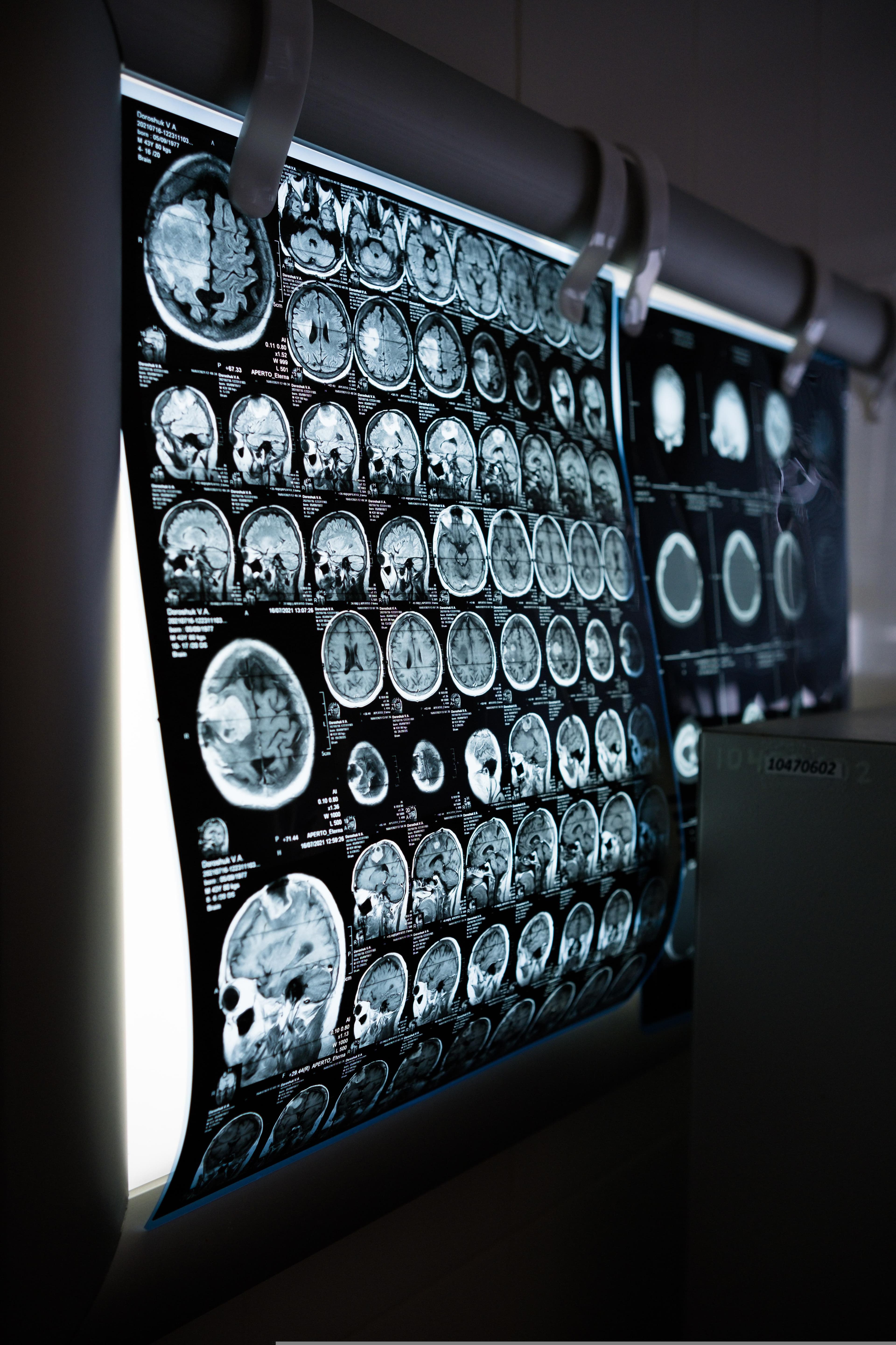
The final lesson of this week’s papers is the most practical. A trauma CT is a photograph taken at one moment, read under pressure and often out of hours. The Emergency Radiology review published on 31 October addresses exactly this problem [2]. The classic blind spot is the bowel and mesentery: a 2023 review in the Korean Journal of Radiology reports that up to 40% of surgically confirmed bowel and mesenteric injuries were not reported prospectively on CT [20]. These injuries look subtle early, the abdomen of an intubated or intoxicated patient tells you little, and the consequences of delay tend to declare themselves a day later on the ward.
That is why this week’s ESR recommendations place as much weight on process as on pixels: radiologists embedded in the trauma multidisciplinary team, and structured, checklist-driven reporting with defined communication pathways [1]. The RCR expects a provisional primary survey report to reach the trauma team leader as soon as possible (within ten minutes of images becoming available when the radiologist is reporting remotely) and a final formal report within an hour [10]. It is also why serial examination and the tertiary survey still matter in a hospital that scans everyone. The scanner does not replace re-evaluation; it changes what you are re-evaluating.
What this means for your ATLS exam
The ATLS written test examines ATLS doctrine, not the practice of the best-equipped trauma centre in Europe. Where the two diverge, answer as the manual would. Five principles will carry you through most imaging questions.
CT is an adjunct to the secondary survey. Chest and pelvic radiographs and FAST or eFAST are the imaging adjuncts to the primary survey. An option that sends a patient to CT before the primary survey is complete and resuscitation is under way is almost certainly wrong.
Haemodynamically abnormal patients do not go to the scanner. A hypotensive patient who has not responded to initial resuscitation and has a positive FAST needs operative haemorrhage control, not “CT to delineate the injuries”.
Response to resuscitation decides the next step. Rapid responders can proceed to CT and further assessment; transient and non-responders need the source found and controlled. This is exactly where ATLS overlaps with the European radiologists’ “stabilisable” patient.
Never delay transfer for imaging. If a patient needs care your hospital cannot provide, ATLS teaches that diagnostic studies must not hold up transfer.
- A negative CT does not end the assessment. Bowel, mesenteric, pancreatic and diaphragmatic injuries can all be missed. Serial examination and re-evaluation remain mandatory, especially when the patient cannot give you a reliable abdominal examination.
Remember the special populations too. In children, minimise radiation by scanning the regions you need rather than everything. In pregnancy, the mother’s assessment comes first, and imaging she needs should not be withheld out of concern for the fetus.
References
1. Limaye N, Masraf H, Rostampour S, Macri F, Iacobellis F, Mueller-Peltzer K, de la Hoz Polo M, Dick E. ESR Essentials: trauma whole-body CT imaging — practice recommendations by the European Society of Emergency Radiology. Eur Radiol. Published online 30 October 2026. doi: 10.1007/s00330-026-12974-x
2. Verma P, Sureka B, Rodha MS. Missed abdominal injuries on initial trauma CT: pitfalls, mimickers and practical pearls. Emerg Radiol. Published online 31 October 2026. doi: 10.1007/s10140-026-02562-3
3. American College of Surgeons Committee on Trauma. Advanced Trauma Life Support Student Course Manual. 11th ed. Chicago: American College of Surgeons; 2025. facs.org — Advanced Trauma Life Support
4. Hanzalova I, Bourgeat M, Demartines N, Ageron FX, Zingg T. The use of whole body computed tomography does not lead to increased 24-h mortality in severely injured patients in circulatory shock. Sci Rep. 2024;14:2169. doi: 10.1038/s41598-024-52657-5
5. The Skeptics’ Guide to Emergency Medicine. SGEM Xtra: This one goes to 11 — ATLS 11th edition. March 2026. thesgem.com
6. Chan O. Primary computed tomography survey for major trauma. Br J Surg. 2009;96(12):1377–1378. doi: 10.1002/bjs.6881
7. Huber-Wagner S, Lefering R, Qvick LM, Körner M, Kay MV, Pfeifer KJ, Reiser M, Mutschler W, Kanz KG; Working Group on Polytrauma of the German Trauma Society. Effect of whole-body CT during trauma resuscitation on survival: a retrospective, multicentre study. Lancet. 2009;373(9673):1455–1461. doi: 10.1016/S0140-6736(09)60232-4
8. Caputo ND, Stahmer C, Lim G, Shah K. Whole-body computed tomographic scanning leads to better survival as opposed to selective scanning in trauma patients: a systematic review and meta-analysis. J Trauma Acute Care Surg. 2014;77(4):534–539. doi: 10.1097/TA.0000000000000414
9. National Institute for Health and Care Excellence. Major trauma: assessment and initial management (NG39). London: NICE; 2016. nice.org.uk/guidance/ng39
10. The Royal College of Radiologists. Major adult trauma radiology guidance. London: RCR; June 2024. rcr.ac.uk (PDF)
11. Sierink JC, Treskes K, Edwards MJ, Beuker BJ, den Hartog D, Hohmann J, Dijkgraaf MG, Luitse JS, Beenen LF, Hollmann MW, Goslings JC; REACT-2 study group. Immediate total-body CT scanning versus conventional imaging and selective CT scanning in patients with severe trauma (REACT-2): a randomised controlled trial. Lancet. 2016;388(10045):673–683. doi: 10.1016/S0140-6736(16)30932-1
12. The Bottom Line. REACT-2: immediate total-body CT scanning versus conventional imaging and selective CT scanning in patients with severe trauma (critical appraisal summary). thebottomline.org.uk
13. Huber-Wagner S, Biberthaler P, Häberle S, Wierer M, Dobritz M, Rummeny E, van Griensven M, Kanz KG, Lefering R; TraumaRegister DGU. Whole-body CT in haemodynamically unstable severely injured patients — a retrospective, multicentre study. PLoS One. 2013;8(7):e68880. doi: 10.1371/journal.pone.0068880
14. Huber-Wagner S, Mand C, Ruchholtz S, Kühne CA, Holzapfel K, Kanz KG, van Griensven M, Biberthaler P, Lefering R. Effect of the localisation of the CT scanner during trauma resuscitation on survival — a retrospective, multicentre study. Injury. 2014;45 Suppl 3:S76–S82. doi: 10.1016/j.injury.2014.08.022
15. Kinoshita T, Yamakawa K, Matsuda H, et al. The survival benefit of a novel trauma workflow that includes immediate whole-body computed tomography, surgery, and interventional radiology, all in one trauma resuscitation room: a retrospective historical control study. Ann Surg. 2019;269(2):370–376. doi: 10.1097/SLA.0000000000002527
16. Bulum A, Peynircioglu B, Filippiadis DK, Pereira PL, Wolf F. ESR Essentials: trauma team and the role of interventional radiology — practice recommendations by the Cardiovascular and Interventional Radiological Society of Europe. Eur Radiol. 2026;36(7):5907–5915. doi: 10.1007/s00330-026-12440-8
17. Pearce MS, Salotti JA, Little MP, et al. Radiation exposure from CT scans in childhood and subsequent risk of leukaemia and brain tumours: a retrospective cohort study. Lancet. 2012;380(9840):499–505. doi: 10.1016/S0140-6736(12)60815-0
18. Nagasawa K, Nihashi T, Iwata M, Terasawa T. Prevalence of incidental findings requiring interventions in CT for trauma patients in the emergency department: a systematic review and meta-analysis. BMJ Open. 2026;16(4):e103045. doi: 10.1136/bmjopen-2025-103045
19. Munk MD, Peitzman AB, Hostler DP, Wolfson AB. Frequency and follow-up of incidental findings on trauma computed tomography scans: experience at a level one trauma center. J Emerg Med. 2010;38(3):346–350. doi: 10.1016/j.jemermed.2008.01.021
20. Kaewlai R, Chatpuwaphat J, Maitriwong W, Wongwaisayawan S, Shin CI, Lee CW. Radiologic imaging of traumatic bowel and mesenteric injuries: a comprehensive up-to-date review. Korean J Radiol. 2023;24(5):406–423. doi: 10.3348/kjr.2022.0998